Seeing Is Believing: The Benefits of Ultrasound for Patients and Providers
Echocardiogram 101: What the Sonographer Is Actually Measuring
Echocardiogram 101: What the Sonographer Is Actually Measuring
An echocardiogram (often just called an echo) is a heart ultrasound. It’s one of the most common, most useful tests in medicine because it doesn’t just take a picture of the heart. It measures how the heart is built and how blood is moving through it.
An echocardiogram (echo) is an ultrasound of your heart. It shows how your heart looks and how it works while it beats.
During the test, the sonographer isn’t just “taking pictures.” They’re collecting measurements so the doctor can answer questions like:
Is the heart pumping well?
Are the heart valves working properly?
Is blood flowing the right way?
Is there too much pressure on the heart or lungs?
Here’s what they’re actually measuring using plain words.

1) The size of the heart chambers
Your heart has 4 main sections (called chambers). The echo checks if any are too big.
Why this matters:
A chamber can get bigger if the heart is working too hard.
Some valve problems can also make chambers enlarge.
2) The thickness of the heart muscle
The echo checks if the heart walls are too thick.
Why this matters:
High blood pressure can make the heart muscle thicker over time.
A thick heart can have trouble relaxing and filling with blood.
3) How strong the heart pumps
This is a big one.
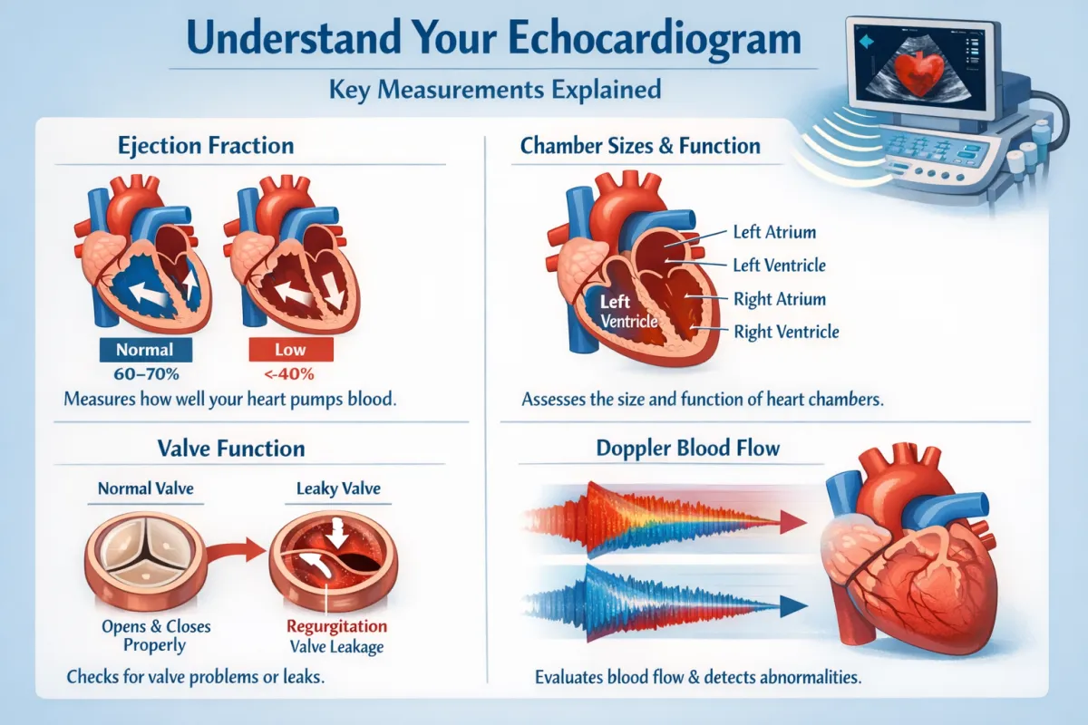
Ejection Fraction (EF)
EF is a number that shows how much blood the heart pumps out with each beat.
If EF is normal, the pumping strength is usually good.
If EF is low, the heart may not be pumping strongly enough.
Important: You can still have symptoms even if EF is normal because pumping is only one part of heart health.
4) How well the heart relaxes and fills
Your heart has to squeeze and also relax.
Sometimes the heart becomes stiff and doesn’t relax well. This can cause symptoms like:
shortness of breath
tiredness
swelling (in some cases)
The echo checks how blood fills the heart and whether the heart is stiff.
5) How the heart valves are working
Your heart has valves that act like doors. The echo checks two main things:
A) Is the valve too tight? (narrowing)
If a valve doesn’t open well, blood has trouble getting through.
B) Is the valve leaking?
If a valve doesn’t close well, blood can leak backward.
Small leaks are very common and often not a big problem. Bigger leaks matter more.
6) Blood flow speed and direction
The echo uses something called Doppler to measure how fast blood is moving and which direction it’s going.
This helps find:
blocked or narrowed valves
leaking valves
pressure problems
7) Pressure on the right side of the heart and lungs
The echo can estimate if there might be high pressure in the lungs (pulmonary pressure). It’s not a perfect measurement, but it gives clues.
This matters because high lung pressure can strain the right side of the heart.
8) How well the right side of the heart is working
The right side pumps blood to the lungs. The echo checks if it’s pumping normally, especially if there are lung or pressure issues.
9) Fluid around the heart
Sometimes fluid builds up around the heart. The echo can detect it and show whether it looks small or large.
What this means in real life
The sonographer’s job is to collect clear images and accurate numbers. The doctor uses those measurements to decide:
whether your heart looks normal
what might be causing symptoms
if you need treatment or follow-up
Simple questions to ask after your echo
Is my heart pumping normally?
Are my valves okay?
Is anything enlarged or thickened?
Is the pressure on my heart or lungs normal?
Do I need follow-up?
For those seeking expert ultrasound services, Atlanta Ultrasound offers quick, efficient, and comprehensive scans. Our team of skilled professionals is dedicated to providing you with the clarity and care you need.
Contact us today to schedule your ultrasound scan and take a decisive step towards understanding your health.
📍 Multiple locations in Metro Atlanta, GA
📞 Contact: 678-590-3300
🌐 Website:www.atlantaultrasound.com
Disclaimer: The content of this blog post, authored by a sonographer, is provided for educational and informational purposes only. It is not intended as medical advice, nor should it substitute for professional medical consultation, diagnosis, or treatment. Always seek the advice of your physician or other qualified health providers with any questions you may have regarding a medical condition or health concerns.