Seeing Is Believing: The Benefits of Ultrasound for Patients and Providers
Breast Ultrasound After Breastfeeding What changes are common (and when to get checked
Breast Ultrasound After Breastfeeding What changes are common (and when to get checked)
After you stop breastfeeding, your breasts don’t instantly snap back to their pre-pregnancy state. They go through a gradual resetmilk production winds down, ducts shrink, glandular tissue settles, and the whole system slowly reorganizes.
That transition is exactly why some people notice lumps, tenderness, or “weird” texture changes after weaning and why a breast ultrasound can look different in someone who recently breastfed compared to someone who never has.
This guide explains what’s commonly seen on ultrasound after breastfeeding, what tends to be harmless, and what deserves a closer look.
Educational only not a diagnosis. If you feel a new or persistent lump, talk to a clinician.

Why breasts can feel different after you wean
During pregnancy and lactation, hormones drive major structural changes: ducts branch and expand, lobules grow, and blood flow increases. The lactating breast can show distended lobules and secretory ducts filled with milk.
When breastfeeding stops, those changes don’t vanish overnight. For a while, you may still have:
leftover milk in ducts
temporarily enlarged ducts
thicker glandular tissue than usual
areas of firmness as the breast “involutes” (returns toward baseline)
That’s normal biology—not your body “acting up.”
Is breast ultrasound safe after breastfeeding?
Yes. Breast ultrasound is considered safe during pregnancy and lactation because it does not use ionizing radiation.
What a breast ultrasound is actually looking at
Ultrasound helps clinicians evaluate things like:
a specific lump you can feel
focal pain (pain in one spot)
certain types of nipple discharge
In lactating or recently lactating patients, ultrasound is often the first imaging test used for a palpable concern or persistent bloody nipple discharge.
Common (often benign) ultrasound findings after breastfeeding
1) Bigger or more visible milk ducts
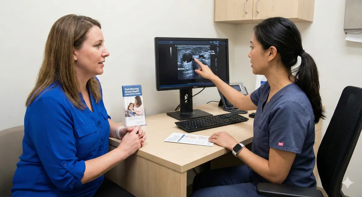
Hormonal and lactation-related changes can cause ductal enlargement (ductal ectasia). One review describes the sonographic appearance as larger ducts/lobules and overall background changes in echogenicity (how bright tissue looks on ultrasound).
What it can feel like: ropy texture, fullness, or mild tenderness especially soon after weaning.
2) Galactocele (milk cyst)
This is one of the most classic “post-breastfeeding” findings.
Galactoceles are described as the most common benign lesion in lactating or recently lactating women, often occurring shortly after stopping breastfeeding due to stagnant milk or duct blockage.
How it can show up:
usually painless, slow-growing lump
ultrasound appearance can vary (sometimes simple and cyst-like, sometimes more complex)
3) Lactating adenoma (a benign tumor that often regresses)
Lactating adenomas commonly appear late in pregnancy or early postpartum and often shrink after lactation ends. Imaging typically looks benign (smooth, circumscribed mass), but there can be overlap with fibroadenomas.
Why this matters: it can feel alarming, but many of these behave benignly—your clinician may monitor or investigate depending on how it looks and whether it changes.
4) Fibroadenoma that “acts different” postpartum
Fibroadenomas can show secretory changes or increased vascularity around pregnancy/lactation, and in some cases can look more complex sometimes prompting biopsy if features look suspicious or the lump grows.
5) Mastitis or abscess-related changes (if you had infection)
If you had mastitis while breastfeeding or right after stopping ultrasound can help evaluate inflammation or an abscess (a pocket of infection). Classic features can include skin thickening and increased local blood flow in inflamed areas.
Important: infection symptoms (redness, warmth, fever, worsening pain) should be treated promptly.

When should I worry? Use this rule
Don’t panic about change. Do take persistence seriously.
Contact a clinician if you have:
a new lump that doesn’t improve over ~1–2 weeks (or grows)
redness, fever, or significant localized warmth/pain
nipple discharge that is persistent, especially bloody (even though it can sometimes resolve on its own, persistence should be evaluated)
skin dimpling, nipple inversion that’s new, or a clearly enlarging mass
Also: imaging of symptoms shouldn’t be delayed just because you recently breastfed—postpartum breast tissue can be trickier to examine, not “off-limits.”
How to prepare for a breast ultrasound after breastfeeding
A simple comfort + image-quality tip: many imaging guidelines recommend nursing or expressing milk before breast imaging because lactating breast tissue can be thicker; emptying can help.
Even after weaning, if you still have some milk production, expressing a little beforehand may reduce tenderness during the exam.
Bottom line
After breastfeeding, it’s common for ultrasound to reflect “recent lactation biology”—visible ducts, cysts like galactoceles, and benign masses that often stabilize or regress.
But here’s the grown-up truth: Common is not the same as ignore it. If something is persistent, changing, painful, or worrying you, get it evaluated. That’s what imaging is for.
For those seeking expert ultrasound services, Atlanta Ultrasound offers quick, efficient, and comprehensive scans. Our team of skilled professionals is dedicated to providing you with the clarity and care you need.
Contact us today to schedule your ultrasound scan and take a decisive step towards understanding your health.
📍 Multiple locations in Metro Atlanta, GA
📞 Contact: 678-590-3300
🌐 Website:www.atlantaultrasound.com
Disclaimer: The content of this blog post, authored by a sonographer, is provided for educational and informational purposes only. It is not intended as medical advice, nor should it substitute for professional medical consultation, diagnosis, or treatment. Always seek the advice of your physician or other qualified health providers with any questions you may have regarding a medical condition or health concerns.